*Info & Image Sources: Harry’s Cosmeticology. Click to enlarge*
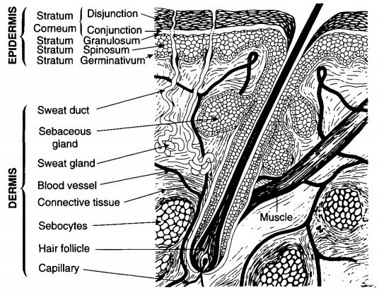
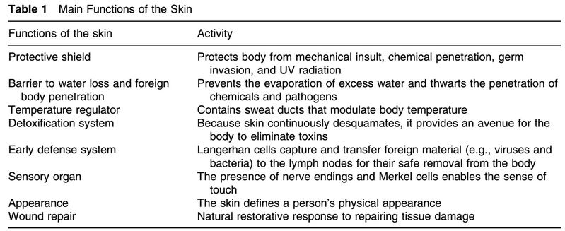
Skin is the organ that protect us from the environment. It is our widest organ with a weight of about 4 kg and 1.8 m2 surface. It prevents dehydration, stop harmful materials from penetrating into our body, helps maintain our body temperature, and act as a cushions against mechanical shock. Our skin is a living organism, therefore it goes through an aging process and under constant renewal. As we aged, its functionality also decreased.
(Info & Image Source: Handbook of Cosmetic Science and Technology. Click to enlarge)
There are 3 layers that make up our skin: The epidermis (outermost layer), the dermis and the subcutis (or the hypodermis).
I’ll be focus most of my research on the epidermis since that’s what our skincare products are trying to target.
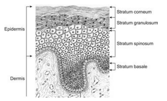
The epidermis is made up primarily of keratinocytes. Keratinocytes produces filamentous protein, keratin, and serves as a protective barrier when combine with different lipid components of the skin.
The epidermis has several layers:
- The basal layer (stratum germinativum) is the innermost
- The stratum spinosum (prickle layer)
- The granulosum layer
- The horny layer (it’s the real name…I did not make it up :D) or the stratum corneum. Outer most layers.
(Info & Image Source: Handbook of Cosmetic Science and Technology. Click to enlarge)
The keratinocytes are form by mitosis and as they matures they move outward from the basal layer and shed on the skin surface. When those cells reach the stratum corneum , the top layer of skin, the cells are called corneocytes and are no longer viable. They lack a nucleus and cellular structures. Corneocytes are flat and filled with water-retaining keratin proteins surrounded by a protein envelope and lipids. There are about 10-30 layers of stacked corneocytes. The thicker skin in our soles and palms has the most layers of stacked corneocytes. These cells remain connected to each other by desmosomes, protein bridges. Stacked bilayers of lipids surround the cells in the extracellular space. This structure is the natural physical and water-retaining barrier of the skin.
The epidermis also contain other cell types: melanocytes (they makes skin pigment), Langerhans cells (colorless), and Merkel cells (relates to sensation). The melanocytes and the Merkel cells are located mainly in the basal layer while the Langerhans cells are distributes throughout.
As the cells mature and move towards the stratum corneum begin to clump protein into granules. These granules are present in the granular cell layer of the skin and are filled with a protein called filaggrin. Filaggrin paired with keratin proteins in the granular cells. This protects filaggrin from proteolytic breakdown into smaller polypeptides or amino acids. But as the degenerating cells move towards the outer layer of the skin toward the stratum corneum, enzymes break down the keratin-filaggrin complex. Filaggrin is on the outside of the corneocytes and water-retaining keratin remains inside the corneocytes of the stratum corneum. When the moisture content of the skin is decreased, specific proteolytic enzymes in the stratum corneum are triggered to further break down filaggrin into free amino acids.
Want to learn more about these amino acids? Continue to Natural Moisturizing Factors (NMF): How our skin retain water naturally